 Euro
Euro
 British Pound
 US Dollar


Product Description
Mouse Anti S-AdenosylHomocysteine (SAH) Clone 301-10 | MA00302-50 | Arthus Biosystems
Product name
Mouse anti-SAH 2a
Catalog Number
MA00302-50
Description
Mouse monoclonal antibody against S-Adenosylhomocysteine [301-10]
Specificity
MA00302 shows the following reactivities with related compounds: S-Adenosylhomocysteine: 100%, S-Adenosylmethionine: -1.5%, Adenosine: <1 %, Homocysteine: < 1%, L-Cysteine: < 1%, Glutathione: < 1%, L-Cystathionine: < 1%, Methythioadenosine (MTA): < 5%, ADP (adenosine diphosphate): < 1%, ATP (adenosine triphosphate): < 1%.
Immunogen
S-Adenosylhomocysteine conjugated to BSA
Properties
Form
Liquid
Storage instructions
Store at 4°C, -20°C for long term storage
Storage buffer
PBS 10mM pH7.4 (NaCI 150mM), Sodium azide 0.02%, BSA 10mg/m1 or PBS 10mM, pH7.4 (NaCI 150mM), Sodium azide 0.02%, Glycerol 50%, BSA 10mg/mi
Purity
>95% Purified from mouse ascites fluid by affinity chromatography
Clonality
Monoclonal
Clone number
301-10
Immunoglobin isotype
IgG3
Affinity
Ka = 4.20 x 10⁸L/mol ( 1.93 x 10⁻⁸M )
Research Areas
- Methylation of biomolecules (DNA, RNA, proteins, hormones, neurotransmitters, etc.)
- One-carbon metabolism
- Signal Transduction
- Metabolism
- Pathways and Processes Cancers
- Arthritis
- Heart diseases
- Neurodegenerative diseases
- Atherosclerosis
- Liver diseases
- Kidney diseases
Applications
The use of MA00302 in the following tested applications has been tested. The application notes include recommended starting dilutions. Optimal dilutions/concentrations should be determined by the end user. Higher dilution than suggested maybe used in IHC and IF. The product may be used in other not-yet-tested applications.
Notes
- cELISA: 1:2000/4000
- FCM : 1:100
- IHC : 1:100
Target
S-adenosylhomocysteine is a competitive inhibitor of S-adenosylmethionine-dependant methyl transferase reactions. Therefore, it plays a key role in the control of methylation via regulation of the intracellular concentration of S-adenosylhomocysteine.
Cellular localization
Cytoplasm, nuclear
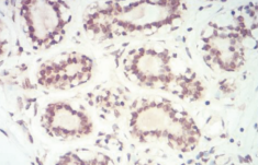
Figure 1: Immunohistochemistry staining was performed using MA00302 with benign breast tissue adjacent to breast cancer. Brown areas indicated positive staining in nuclear and cytoplasmic areas (x400).
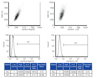
Figure 2: Control for FCM. Normal liver cells L02 and carcinoma cells Hep G2 were stained with the buffer without any antibody.

Figure 3: FCM results from normal liver cell line L02 and hepatocyte carcinoma cell line Hep G2 stained with anti-SAH monoclonal antibody from clone 301-10. Average fluorescence signal in Hep G2 cells (56.99) was reduced compared to that in L02 cells (103.36), indicating SAM level is reduced during carcinogenesis.