Product Description
Mouse Anti S-AdenosylHomocysteine (SAH) Clone 844-1 | MA00305 | Arthus Biosystems
Product name
Mouse anti-SAH 5a/b
Catalog Number
MA00305-50/100
Description
Mouse monoclonal antibody against S-Adenosylhomocysteine [844-1]
Specificity
MA00305 shows the following reactivities with related compounds: S-Adenosylhomocysteine: 100%, S-Adenosylmethionine: -2.6%, Adenosine: < 1%, Homocysteine: < 1%, L-Cysteine: < 1%, Glutathione: < 1%, L-Cystathionine: < 1%, Methythioadenosine (MTA): < 5%, ADP (adenosine diphosphate): < 1%, ATP (adenosine triphosphate): < 1%.
Immunogen
S-Adenosylhomocysteine conjugated to BSA
Properties
Form
Liquid
Storage instructions
Store at 4°C, -20°C for long term storage
Storage buffer
PBS 10mM pH7.4 (NaCI 150mM), Sodium azide 0.02%, BSA 10mg/m1 or PBS 10mM, pH7.4 (NaCI 150mM), Sodium azide 0.02%, Glycerol 50%, BSA 10mg/m1
Purity
>95% Purified from mouse ascites fluid by affinity chromatography
Clonality
Monoclonal
Clone number
844-1
Immunoglobin isotype
IgG2a
Affinity
Ka = 7.05 x 10⁹L/mol( 1.42 x 10⁻¹⁹M )
Research Areas
- Methylation of biomolecules (DNA, RNA, proteins, hormones, neurotransmitters, etc.)
- One-carbon metabolism
- Signal Transduction
- Metabolism
- Pathways and Processes Cancers
- Arthritis
- Heart diseases
- Neurodegenerative diseases Atherosclerosis
- Liver diseases
- Kidney diseases
Applications
The use of MA00305 in the following tested applications has been tested. The application notes include recommended starting dilutions. Optimal dilutions/concentrations should be determined by the end user. Higher dilution than suggested maybe used in IHC and IF. The product may be used in other not-yet-tested applications.
Notes
- cELISA: 1:30,000/36,000
- FCM: 1:300
- IHC: 1:300
Target
S-adenosylhomocysteine is a competitive inhibitor of S-adenosylmethionine-dependant methyl transferase reactions. Therefore, it plays a key role in the control of methylation via regulation of the intracellular concentration of S-adenosylhomocysteine.
Cellular localization
Cytoplasm, nuclear

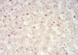
Figure 1: Immunohistochemistry staining was performed using MA00305 with benign liver tissue adjacent to liver cancer. Brown areas indicated strong positive staining in nuclear and cytoplasmic areas (x400).
 Euro
Euro
 British Pound
 US Dollar

